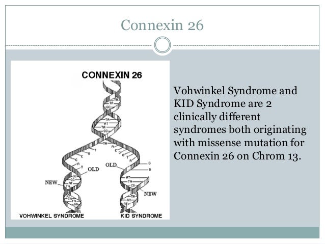
Sunday, March 29, 2015

Cell Biology


                                                 Vohwinkel syndrome

            vohwinkel syndrome is a disorder with classic and variant forms, both of which affect the skin.
             The classic form of Vohwinkel syndrome is caused by mutations in the GJB2 gene. This gene provides instructions for making a protein called gap junction beta 2, more commonly known as connexin 26. Connexin 26 is a member of the connexin protein family. Connexin proteins form channels called gap junctions that permit the transport of nutrients, charged atoms (ions), and signaling molecules between neighboring cells that are in contact with each other. Gap junctions made with connexin 26 transport potassium ions and certain small molecules.
           Connexin 26 is found in cells throughout the body, including the inner ear and the skin. In the inner ear, channels made from connexin 26 are found in a snail-shaped structure called the cochlea. These channels may help to maintain the proper level of potassium ions required for the conversion of sound waves to electrical nerve impulses. This conversion is essential for normal hearing. In addition, connexin 26 may be involved in the maturation of certain cells in the cochlea. Connexin 26 also plays a role in the growth, maturation, and stability of the outermost layer of skin (the epidermis).
             The GJB2 gene mutations that cause Vohwinkel syndrome change single protein building blocks (amino acids) in connexin 26. The altered protein probably disrupts the function of normal connexin 26 in cells, and may interfere with the function of other connexin proteins. This disruption could affect skin growth and also impair hearing by disturbing the conversion of sound waves to nerve impulses.


Cell Biology


                                               Charcot-Marie-Tooth disease

                Charcot-Marie-Tooth disease (CMT) is one of the most common inherited neurological disorders, affecting approximately 1 in 2,500 people in the United States.
The hypothesis is that CMTX mutations lead to a loss of normal cellular communication, which in turn may lead to Schwann cell dysfunction and peripheral neuropathy.

             Mutations in Cx32 may give rise to CMTX through one of several proposed mechanisms. Some mutations will lead to loss of function with no possibility of expression of functional channels. One non-sense mutation (ARG22STOP) is at a very upstream site and would likely lead to the expression of no functional protein. Other mutations in Cx32 lead to the abnormal accumulation of Cx32 proteins in the cytoplasm, particularly in the Golgi apparatus, and in many of those cases Cx32 is not identified in the plasma membrane.
              Cx32 forms a gap junction channels that allows the passage of molecules smaller than ~7 Å in radius.  Second messengers like cAMP and Ca2+ permeate wild-type Cx32 junctions.  As with all connexins, the complete cell-cell channel is composed of two opposing hemichannels, called connexons, each anchored in the plasma membrane of the opposing cells. 
                  Another very common result from the mutation that is shown in every CMT disease is the demyelination around neuron's axon.  The axon forms the connection between the Central Nervous System (the brain), and the Peripheral Nervous System (extremities), which send information back and forth.  The myelin sheath surrounds the axon and acts to produce an insulation, allowing the message known as action potential flow through faster.  People with CMTX lack the myelin sheath, resulting in slower messages from the brain to the extremities and muscles.

Picture


Microbioogy


                                           Legionella Disease

Legionnaires' disease is a severe form of pneumonia — lung inflammation usually caused by infection. Legionnaires' disease is caused by Gram-negative aerobic bacteria known as legionella.
You can't catch Legionnaires' disease from person-to-person contact. Instead, most people get Legionnaires' disease from inhaling the bacteria. Older adults, smokers and people with weakened immune systems are particularly susceptible to Legionnaires' disease.
These species cause a less severe infection known as Pontiac fever, which resembles acute influenza. These bacterial species can be water-borne or present in soil. Patients with Legionnaires' disease usually have fever, chills, and a cough, which may be dry or may produce sputum. Some patients also have muscle aches, headache, and tiredness, loss of appetite, loss of coordination (ataxia), and occasionally diarrhea and vomiting.


Microbiology


                                                          West Nile virus (WNV)

West Nile virus (WNV) is an infectious disease that first appeared in the United States in 1999. Infected mosquitoes spread the virus that causes it. People who get WNV usually have no symptoms or mild symptoms. The symptoms include a fever, headache, body aches, skin rash, and swollen lymph glands. They can last a few days to several weeks, and usually go away on their own.
If West Nile virus enters the brain, however, it can be life-threatening. It may cause inflammation of the brain, called encephalitis, or inflammation of the tissue that surrounds the brain and spinal cord, called meningitis.

West Nile virus (WNV) is transmitted through female mosquitoes, which are the prime vectors of the virus. The mosquito species that are most frequently infected with WNV feed primarily on birds.

File:West Nile virus transmission cycle.jpg


Thursday, March 5, 2015

Cell Biology


                                                               Kinesin
A kinesin is a protein belonging to a class of motor proteins found in eukaryotes cells.
Kinesins move along microtubule (MT) filaments, and are powered by the hydrolysis of ATP, thus kinesins are ATPases. The active movement of kinesins supports several cellular functions including mitoses, meiosis and transport of cellular cargo, such as in axonal transport. Most kinesins walk towards the plus end of a microtubule, which, in most cells, entails transporting cargo from the centre of the cell towards the periphery. This form of transport is known as anterograde transport. In contrast, dyneins are motor proteins that move toward the microtubules' minus end.


Image result for kinesin


                                                    Dynein
Dynein is a motor protein in cells which converts the chemical energy contained in ATP into the mechanical energy of movement. Dynein transports various cellular cargo by "walking" along cytoskeletal microtubules towards the minus-end of the microtubule, which is usually oriented towards the cell center. Thus, they are called "minus-end directed motors." This form of transport is known as retrograde transport. In contrast, kinesins, which are motor proteins that move toward the microtubules' plus end, are called plus-end directed motors.


                                                                      Profilin
Profilin is an actin binding protein involved in the dynamic turnover and restructuring of the actin cytoskeleton. It is found in most of eukaryotes cells. Profilin is important for spatially and temporally controlled growth of actin microfilaments, which is an essential process in cellular locomotion and cell shape changes. This restructuring of the actin cytoskeleton is essential for processes such as organ development, wound healing, and the hunting down of infectious intruders by cells of the immune system.  
Profilin binds some variants of membrane phospholipids. The function of this interaction is the sequestration of profilin in an "inactive" form, from where it can be released by action of the enzyme phospholipase C.
Profilin is the major allergen present in birch, grass, and other pollen.  


Cell Biology


                                                         Leigh syndrome
Leigh's disease is a rare inherited neurometabolic disorder that affects the central nervous system. This progressive disorder begins in infants between the ages of three months and two years. Rarely, it occurs in teenagers and adults. Leigh's disease can be caused by mutations in mitochondrial DNA or by deficiencies of an enzyme called pyruvate dehydrogenase. Symptoms of Leigh's disease usually progress rapidly. The earliest signs may be poor sucking ability,and the loss of head control and motor skills.These symptoms may be accompanied by loss of appetite, vomiting, irritability, continuous crying, and seizures. As the disorder progresses, symptoms may also include generalized weakness, lack of muscle tone, and episodes of lactic acidosis, which can lead to impairment of respiratory and kidney function.

In Leigh’s disease, genetic mutations in mitochondrial DNA interfere with the energy sources that run cells in an area of the brain that plays a role in motor movements. The primary function of mitochondria is to convert the energy in glucose and fatty acids into a substance called adenosine triphosphate (ATP). The energy in ATP drives virtually all of a cell's metabolic functions. Genetic mutations in mitochondrial DNA, therefore, result in a chronic lack of energy in these cells, which in turn affects the central nervous system and causes progressive degeneration of motor functions.
There is also a form of Leigh’s disease (called X-linked Leigh's disease) which is the result of mutations in a gene that produces another group of substances that are important for cell metabolism. This gene is only found on the X chromosome. 
The most common treatment for Leigh's disease is thiamine or Vitamin B1. Oral sodium bicarbonate or sodium citrate may also be prescribed to manage lactic acidosis. Researchers are currently testing dichloroacetate to establish its effectiveness in treating lactic acidosis. In individuals who have the X-linked form of Leigh’s disease, a high-fat, low-carbohydrate diet may be recommended.


leighsdiseaseremediesinfo2


Microbiology


                                      The role of helicobacter pylori in peptic ulcer

Helicobacter pylori bacteria commonly live in the mucous layer that covers and protects tissues that line the stomach and small intestine. Often, H. pylori causes no problems, but it can cause inflammation of the stomach's inner layer, producing an ulcer.

It's not clear how H. pylori spreads. It may be transmitted from person to person by close contact, such as kissing. People may also contract H. pylori through food and water.

After H. pylori enters body, it attacks the lining of stomach, which usually protects from the acid which uses to digest food. Once the bacteria have done enough damage, acid can get through the lining, which leads to ulcers. These may bleed, cause infections, or keep food from moving through digestive tract.

File:H pylori ulcer diagram en.png

Microbiology


                                                            Toxoplasma Gondii


The T. gondii life cycle has three stages: tachyzoite, bradyzoite, and sporozoite. During the acute stage of T. gondii infection, tachyzoites invade and replicate within cells and are responsible for congenital infection. The tachyzoites invade all organs, especially the muscles (including the heart), liver, spleen, lymph nodes, and central nervous system (CNS). During latent infection, bradyzoites are present in tissue cysts. Sporozoites are found in environmentally resistant oocysts formed after the sexual stage of the life cycle.
Cats are the definitive hosts for the sexual stage of T. gondii, which takes place in their intestinal mucosa. During acute infection, cats excrete non-infectious oocysts in their feces. Depending on environmental conditions, the oocysts sporulate and become infectious after one day to several weeks. Under favorable conditions (i.e., in warm, moist soil), oocysts remain infectious for a year or more.
T. gondii is transmitted to humans by three principal routes.
 First, humans can acquire T. gondii by eating raw or inadequately cooked infected meat, especially pork, mutton, and wild game, or uncooked foods that have come in contact with infected meat.
Second, humans can inadvertently ingest oocysts that cats have passed in their feces, either from a litter box or from soil.
Third, women can transmit the infection transplacentally to their unborn fetus. In adults, the incubation period for T. gondii infection ranges from 10 to 23 days after the ingestion of undercooked meat and from five to 20 days after the ingestion of oocysts from cat feces.