Saturday, April 25, 2015

Microbiology


                                         Overview of Hypersensitivity Reactions


·                         Altered reaction to an Ag that results in pathologic reactions upon the exposure of a sensitized host to that Ag
·                         Not necessarily an “over-reactive” immune response, but an inappropriate one (“abnormally active”)
·                         Both endogenous and exogenous Ag may elicit hypersensitivity reactions
·                         The development of hypersensitivity diseases is often associated with the inheritance of particular susceptibility genes. 
·                         All reaction types have a sensitization phase and effector phase
·                         Hypersensitivity reflects an imbalance between the effector mechanisms of immune responses and the control mechanisms that serve to normally limit such responses.
·                         Four types based on the mechanism by which immune reactions initiate tissue injury

·                         Increasingly recognized that multiple mechanisms may be operative in any one hypersensitivity disease and some disease represent a continuum of reaction types.


Type
Prototype Disorder
Immune Mechanisms
Pathologic Lesions
Type I hypersensitivity (immediate)
Anaphylaxis; allergies; bronchial asthma (atopic forms)
Production of IgE antibody → immediate release of vasoactive amines and other mediators from mast cells; recruitment of inflammatory cells (late-phase reaction)
Vascular dilation, edema, smooth muscle contraction, mucus production, inflammation
Type II hypersensitivity
IMHA; Good pasture syndrome
Production of IgG, IgM → binds to antigen on target cell or tissue → phagocytosis or lysis of target cell by activated complement or Fc receptors; recruitment of leukocytes
Cell lysis; inflammation
Type III hypersensitivity
(immune complex- mediated)
SLE; some forms of glomerulonephritis; serum sickness; Arthus reaction
Deposition of antigen-antibody complexes → complement activation → recruitment of leukocytes by complement products and Fc receptors → release of enzymes and other toxic molecules
Necrotizing vasculitis (fibrinoid necrosis); inflammation
Cell-mediated (type IV) hypersensitivity
Contact dermatitis; multiple sclerosis; type I, diabetes; transplant rejection; tuberculosis
Activated T lymphocytes → i) release of cytokines and macrophage activation; ii) T cell-mediated cytotoxicity
Perivascular cellular infiltrates; edema; cell destruction; granuloma formation


Cell biology


                                                         Cell cycle check point

            The cell cycle checkpoints determine if a cell is ready to progress to the next stage.
            Late in the G1 phase, the G1 checkpoint determines if the cell will enter the following S phase.
            In animals, the G1 checkpoint or restriction point, is largely controlled by growth factors.
The G2 checkpoint determines if the cell will enter the M phase and requires the proper completion of DNA synthesis.
         The third cell cycle checkpoint is the spindle assembly checkpoint which occurs between metaphase and anaphase and requires the proper attachment of all the chromosomes to the spindle apparatus.
          The fusion of cells in different stages of the cell cycle (to form a heterokaryon) demonstrated that lat    ter stages possess factor trigger progression. 

G1 + S _____ G1 nucleus enters S phase immediately. 
S + G2 _____  G2 nucleus does not enter S phase before mitosis. 
M+ G1, S or G2  ____non-M phase nuclei enters mitosis, whether or not the DNA is duplicated.


Saturday, April 18, 2015

Cell Biology


                                                               Rho family of GTPases


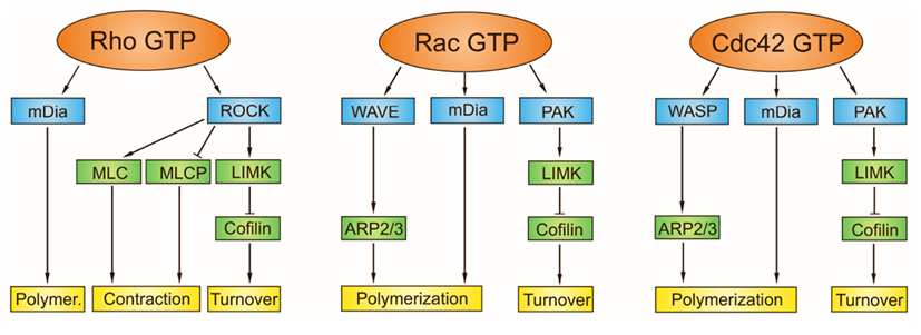
                 The Rho family of GTPases is a family of small (21 kDa) signaling G protein, and is a subfamily of the Ras superfamily. The members of the Rho GTPase family have been shown to regulate many aspects of intracellular actin dynamics, and are found in all eukaryotic kingdoms, including yeasts and some plants. Three members of the family have been studied in detail: Cdc42, Rac1, and Rh0A. All G proteins are "molecular switches", and Rho proteins play a role in organelle development, cytoskeleton dynamics, cell movement, and other common cellular functions.


Friday, April 17, 2015

Cell Biology


                                            Intrinsic apoptotic pathway

         The intrinsic signaling pathway for programmed cell death involves non-receptor–mediated intracellular signals, inducing activities in the mitochondria that initiate apoptosis.
Stimuli for the intrinsic pathway include viral infections or damage to the cell by toxins, free radicals, or radiation. Damage to the cellular DNA can also induce the activation of the intrinsic pathway for programmed cell death. These stimuli induce changes in the inner mitochondrial membrane that result in the loss of transmembrane potential, causing the release of pro-apoptotic proteins into the cytosol.
Pro-apoptotic proteins activate caspases that mediate the destruction of the cell through many pathways. These proteins also translocate into the cellular nucleus, inducing DNA fragmentation, a hallmark of apoptosis.
The regulation of pro-apoptotic events in the mitochondria occurs through activity of members of the Bcl-2 family of proteins and the tumor suppressor protein p53. Members of the Bcl-2 family of proteins may be pro- or anti-apoptotic7
The anti-apoptotic proteins are Bcl-2, Bcl-x, Bcl-xL, Bcl-XS, Bcl-w, and BAG. Some of these proteins are currently under investigation as potential targets for anticancertherapy.
Pro-apoptotic proteins include Bcl-10, Bax, Bak, Bid, Bad, Bim, Bik, and Blk. It has been suggested that upregulation of these proteins or their increased activation may offer an approach for cancer therapy.Cellular pathways that modulate the activities of the p53 protein are also currently being evaluated as targets for potential anticancer therapies.

Elements of the intrinsic apoptosis pathway image 

Cell Biology


                                                               MPF regulation

                       Cyclin-CDK (cyclin-dependent protein kinase) complexes play a central role in cell cycle progression . The function of cyclin-CDKs is to run the cell cycle smoothly, and these are therefore called “cell cycle engines.” Cyclins are proteins that vary in quantity throughout the cell cycle. The cyclins are expressed between the G1 and S phases, during the S phase, and between the G2 and M phases, known as the G1/S-phase cyclin, the S-phase cyclin, and the G2/M-phase cyclin, respectively. Each cyclin is rapidly synthesized during a specific phase of the cell cycle and is again promptly broken down after it serves its purpose. They are broken down not only because they are no longer needed, but because the breakdown is required for the cell cycle to transition to the next step. Meanwhile, the CDKs need to be synthesized de novo when cell proliferation starts from the G0 phase. Once the cell cycle starts, some types of CDK are broken down during a specific phase of the cell cycle, whereas others are not. When the cyclins and CDKs that are expressed in a specific phase are bonded and activated, they phosphorylate the specific serine and threonine residues of a target protein. The phosphorylated target protein executes the events occurring in the respective phases of the cell cycle.

Cell cycle control by cyclin-CDK

Microbiology


                                                               Salmonellosis

                          Salmonella infection is usually caused by eating raw or undercooked meat, poultry, eggs or egg products. The incubation period ranges from several hours to two days. Most salmonella infections can be classified as gastroenteritis. Possible signs and symptoms include:

Nausea
Vomiting
Abdominal cramps
Diarrhea
Fever
Chills
Headache
Blood in the stool
           Signs and symptoms of salmonella infection generally last four to seven days, although it may take several months for your bowels to return to normal.
Salmonella bacteria live in the intestines of people, animals and birds. Most people are infected with salmonella by eating foods that have been contaminated by feces. Commonly infected foods include:
·         Raw meat, poultry and seafood. 
·          Raw eggs. 
·          Fruits and vegetables.  


Persistent bacterial infections: the interface of the pathogen and the host immune system

Microbiology


                                               Hemolytic disease of the newborn


                     Hemolytic disease of the fetus and newborn (HDFN), erythroblastosis fetalis, is a blood disorder that occurs when the blood types of a mother and baby are incompatible.
The mother produces substances called antibodies that attack the developing baby's red blood cells.
                    The most common form of HDN is ABO incompatibility, which is usually not very severe. Other, less common types may cause more severe problems.
                   The least common form is Rh incompatibility, which can almost always be prevented. When this form does occur, it can cause very severe anemia in the baby.
HDN can destroy the newborn baby's blood cells very quickly, which can cause symptoms such as:

                         Edema (swelling under the surface of the skin)
·                                                             Newborn jaundice
    Signs of HDN include:
  • Anemia or low blood count
  • Enlarged liver or spleen
  • Hydrops that can lead to heart failure from too much fluid.

Microbiology


                              Methods of Detection of Hypersensitivity Reactions
 
                  Hypersensitivity reactions are classified into four groups: Type I, II, III, and IV, each         characterized by specific biological actions.
          Type I hypersensitivities can be determined by provacation testing, immediate-type skin testing, or radioallergosorbent tests (RASTs); Type II hypersensitivities can be determined by measuring the level of IgG antibodies to specific host proteins; Type III hypersensitivities may be detected with serum IgG antibody testing to specific antigens; Type IV hypersensitivities are determined by delayed skin testing or memory lymphocyte immunostimulation assay (MELISA).